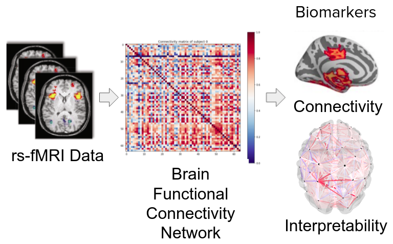
Ayu estimates a person's age from a resting-state fMRI scan, then uses the model's attention to show which brain connections change as we get older.
The problem
People of the same age can have brains that have aged quite differently. If a scan could show how old a brain looks, set against how old its owner really is, that gap could become an early, non-invasive sign of healthy ageing, or of ageing going off track.
Resting-state fMRI is a promising place to look. The person lies still in the scanner and does nothing in particular while the machine records activity across the brain. Regions whose activity rises and falls together are called functionally connected, and the pattern of those connections carries a lot of information about age.
Getting a model to read that pattern is harder than it sounds. Each scan is noisy, and a little head movement can swamp the signal. Brain datasets are small next to the image collections that deep networks are usually trained on, so overfitting is never far away. Earlier work often looked at a single network, such as the default mode network, or used small samples, or spanned wide age gaps that left middle-aged adults thinly represented. And underneath all of it sits an awkward question: to compare brains you first have to cut them into regions, and nobody knows for sure which cut matches how the brain is really organised.
What we built
We worked with 638 healthy adults from the Cam-CAN repository, aged 20 to 88. After standard cleaning of the scans (motion correction, alignment to a common brain template, and removal of signal coming from white matter and cerebrospinal fluid), we divided each brain into regions in two different ways, once with 100 regions and once with 64. For every person we then measured how strongly each pair of regions moves together. That gives a connectivity matrix, which a network can read much as it reads an image.
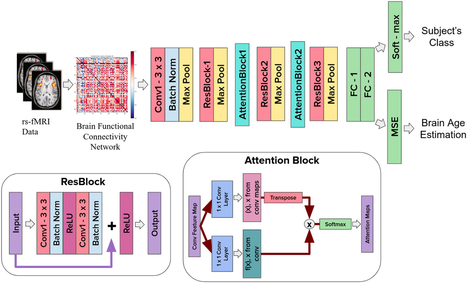
The Ayu pipeline. A connectivity matrix goes into a ResNet with attention blocks, which has two outputs: an age group and an age in years. The two panels underneath show the residual block and the attention block in detail. Figure 1 of the paper.
The network is a small ResNet with attention blocks. The attention lets it concentrate on the connections that matter for age instead of weighing them all equally. Afterwards we used integrated gradients to trace each prediction back to specific connections, so a neuroscientist can see why the model said what it said, and not only what it said.
What we found
- On the 100-region setup, the model sorted people into four age groups with about 72.6 percent accuracy.
- Predicting age directly, it was off by about 6.8 years on average, with an r-squared of 0.75.
- Adding attention improved the results over the same networks without it.
- Connectivity in the left prefrontal cortex, the primary somatosensory cortex and the default mode network fell steadily with age, while early visual regions and the right ventral attention network peaked around middle age.
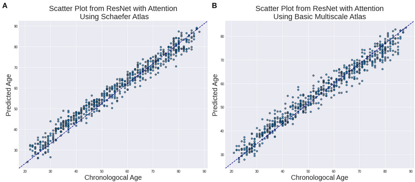
Predicted age against actual age, for the 100-region atlas (left) and the 64-region atlas (right). The closer a point sits to the dashed line, the better the prediction. Figure 2 of the paper.
The maps below show where the model was looking. For each of the four age groups, they mark which brain regions carried the most weight in its decision.
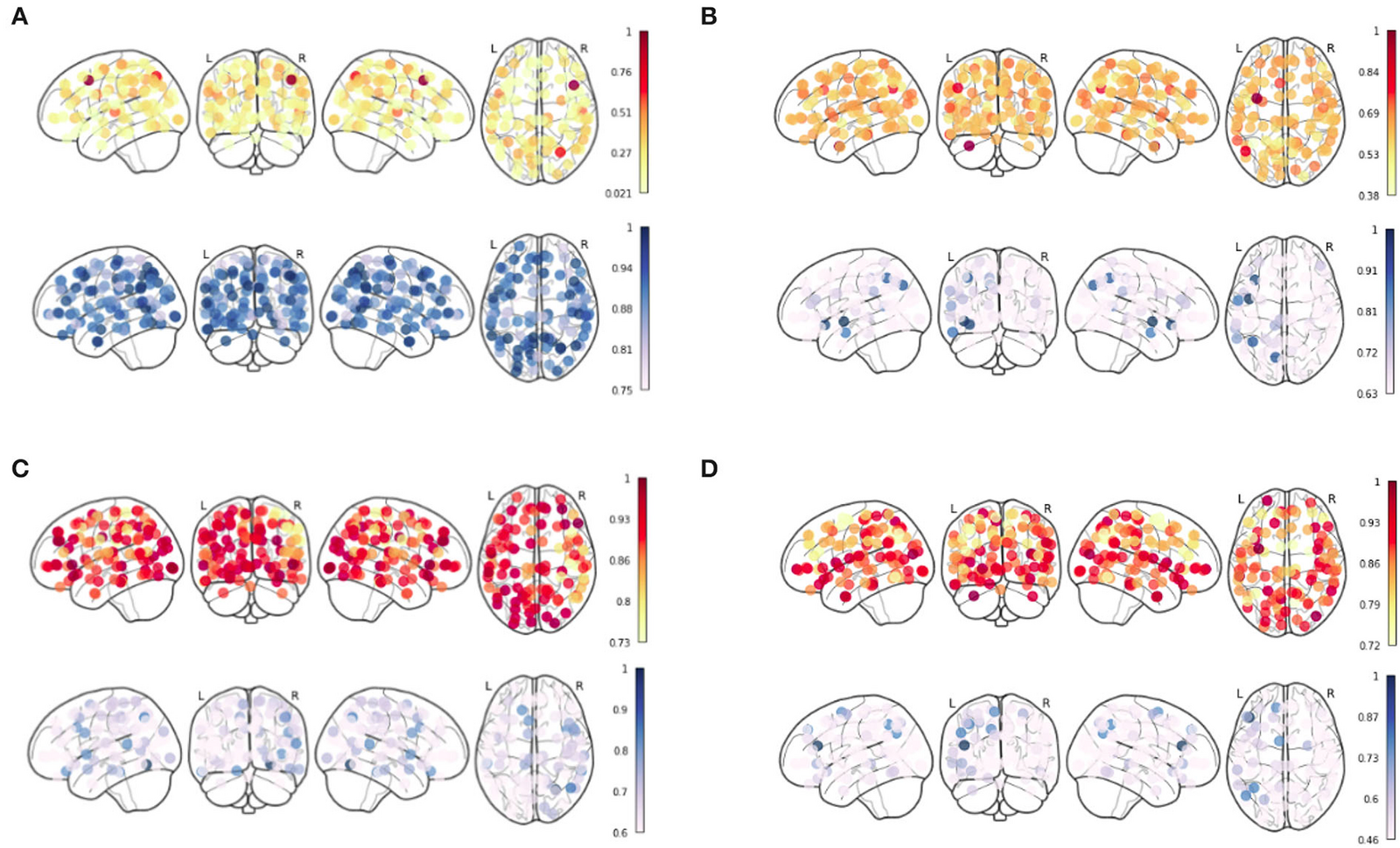
Node strength inferred by the model using integrated gradients, on the 100-region atlas, one panel for each age group. Figure 3 of the paper.
These results come from one dataset and one held-out split, so we treat them as a starting point, not a final answer. They do suggest that whole-brain connectivity holds real information about healthy ageing.
Read the paper
Ayu: Characterization of Healthy Ageing from Neuroimaging Data with Deep Learning and rsfMRI, by Kushal Borkar, Anusha Chaturvedi, Vinod P. K. and Raju Surampudi Bapi, published in Frontiers in Computational Neuroscience, 2022.
The figures above are reproduced from the paper, which is published open access under a Creative Commons Attribution (CC BY) licence.
More work
Back to Works
Interactive Machine Learning
Outlining an organ on a CT or MRI scan means tracing it on a hundred slices or more, one at a time. We built a way to make that far less painful.
Covid-19 Cough Detector
A COVID-19 screening tool that listens to a short cough recording and predicts whether it's positive. Our team placed 7th in the Microsoft Azure AI Challenge.